
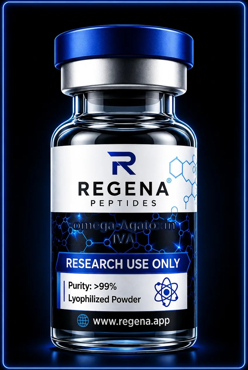
Other · Disulphide-rich voltage-gated calcium-channel gating modifier
omega-Agatoxin IVA
Also known as ω-Agatoxin IVA; ω-Aga-IVA
A 48-residue spider toxin used to resolve CaV2.1-dependent calcium currents through voltage-dependent gating inhibition.
- Length
- 48 amino acids
- Half-life
- Not well characterised
- Mechanism
- omega-Agatoxin IVA interacts with extracellular voltage-sensor elements of CaV2.1 channels and impedes activation by favouring closed states. It inhibits P-type currents with particularly high affinity, while Q-type current sensitivity varies with channel composition and experimental conditions.
- Studied in
- Recombinant CaV2.1 assays, cerebellar Purkinje-cell recordings and isolated neuronal preparations use the peptide to examine P/Q-type calcium currents and presynaptic calcium entry.
About omega-Agatoxin IVA
omega-Agatoxin IVA is a 48-amino-acid peptide isolated from venom of the funnel-web spider Agelenopsis aperta. Four disulphide bonds stabilise its compact structure, which includes a cystine-knot core. Its characterisation provided an important tool for distinguishing P-type calcium currents in cerebellar Purkinje cells from other high-voltage-activated calcium currents. The Roman numeral and letter designation identify a particular agatoxin member and should not be interchanged with those of related venom peptides with different pharmacological profiles.
The principal molecular target is CaV2.1, encoded by CACNA1A, which underlies both P-type and Q-type currents. Differences in splice form, auxiliary subunits and cellular context contribute to the pharmacological variation between these currents. Rather than behaving as a simple pore blocker, omega-Agatoxin IVA modifies voltage-dependent activation, and strong depolarisation can relieve inhibition. Laboratories combine toxin sensitivity with voltage protocols and molecular controls to resolve CaV2.1 contributions within mixed populations of calcium channels.
Laboratory notes
- Record holding and conditioning potentials because strong depolarisation can alter toxin inhibition.
- Report CaV2.1 splice form and auxiliary subunits in recombinant assays wherever known.
- Allow for preparation-dependent equilibration and compare against other calcium-channel probes when analysing mixed currents.
Storage and reconstitution basics: storage guide.
Questions about omega-Agatoxin IVA
Are P-type and Q-type currents produced by different channel genes?
Both are associated with CaV2.1/CACNA1A. Splicing, auxiliary subunits and cellular context contribute to their different electrophysiological and pharmacological properties.
Why can strong depolarisation change inhibition?
The peptide modifies channel gating, so voltage-driven changes in channel conformation can reduce its inhibitory effect.
Is it equivalent to an N-type calcium-channel blocker?
No. Its principal target is CaV2.1, whereas N-type currents are mediated by CaV2.2.
Is omega-Agatoxin IVA for human use?
No. It is supplied strictly for in-vitro laboratory research and is not for human or animal consumption.
Related research peptides
All products are sold for laboratory research use only. Not for human consumption, not a medicine, not for diagnosing, treating or preventing any condition.